
Research on the pediatric head (ages newborn to 18-years-old) at Duke University's Injury and Orthopaedic Biomechanics Laboratory focuses on two major goals: to develop biofidelic child ATDs and to improve finite element models. The research projects currently underway at the lab are the quantitatively evaluating Hybrid III scaling rules that govern the current design of the child ATDs and enhancing the development of accurate FEM models that are used as a substitute for mechanical testing.
The current child ATD heads were developed by scaling adult models. Hybrid III scaling rules are used define the mass, center of gravity, and impact response of the pediatric heads. Our research will experimentally determine these properties for a variety of child ages using both biomechanical testing and computational modeling. This data will be used to conduct a quantitative analysis of the current scaling rules and examine the error associated with using the scaling rules. The lab is developing a new scaling rule that will be used to determine the mass moment of inertia to be used for the pediatric head.
The lab is working to increase the accuracy of finite element models by providing accurate constitutive properties of the sutures and fontanelles and accurate geometry for the pediatric head. At birth, the skull is composed of five separate bones, connected by suture and fontanelle and by age 2, the skull is completely fused into a solid bone. Viscoelastic tensile testing of the pediatric suture and fontanelle is being performed to obtain the tissues? viscoelastic properties, yield stress, yield strain, ultimate stress, and ultimate strain properties.
The lab is exploring thresholding techniques for developing anatomically detailed finite element meshes from high-resolution CT images. Preprocessing of CT DICOM data is done using AMIRA in collaboration with the Duke University Visualization Technology Group. These data are then uploaded to HyperMesh (Altair Engineering) for finite element meshing.
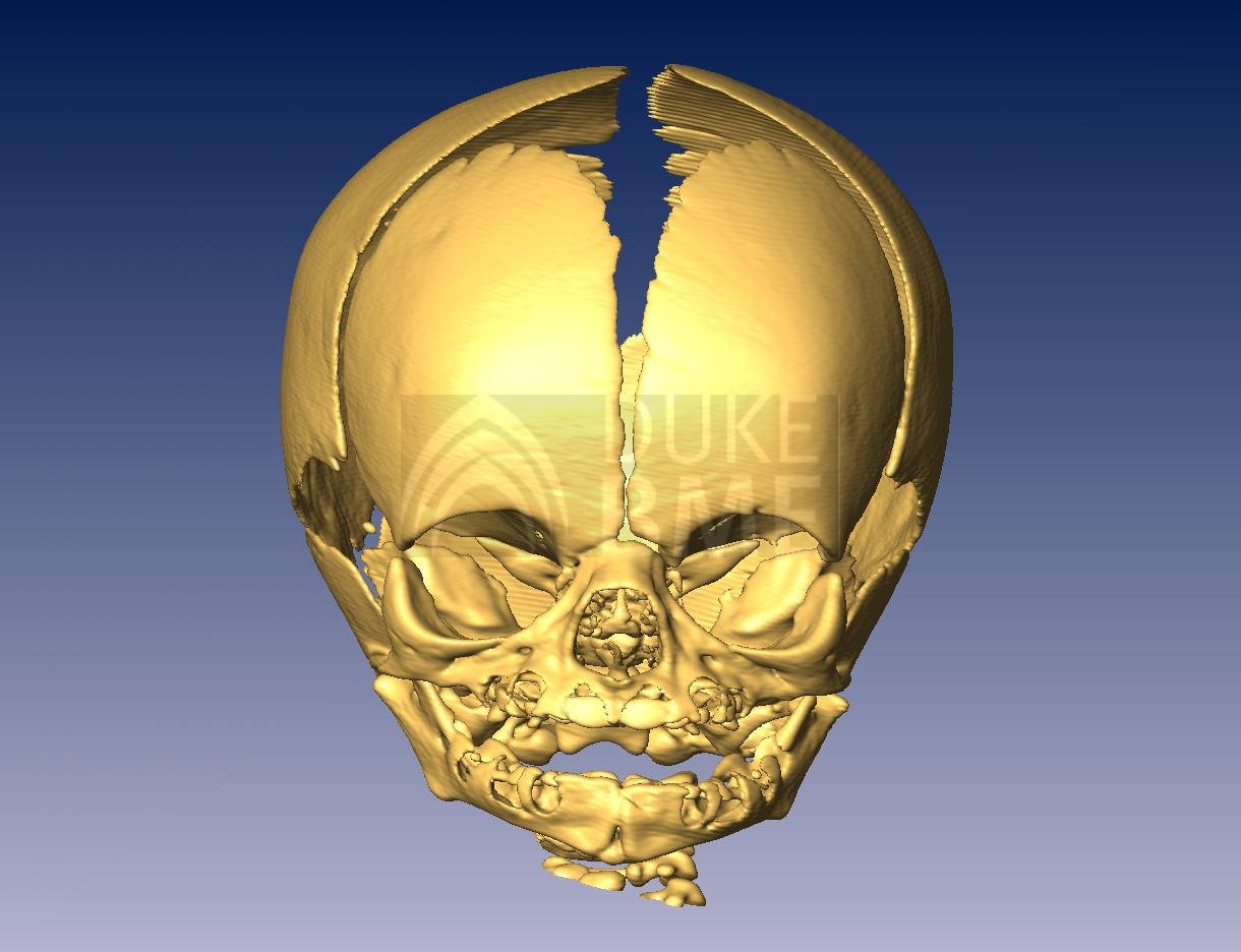
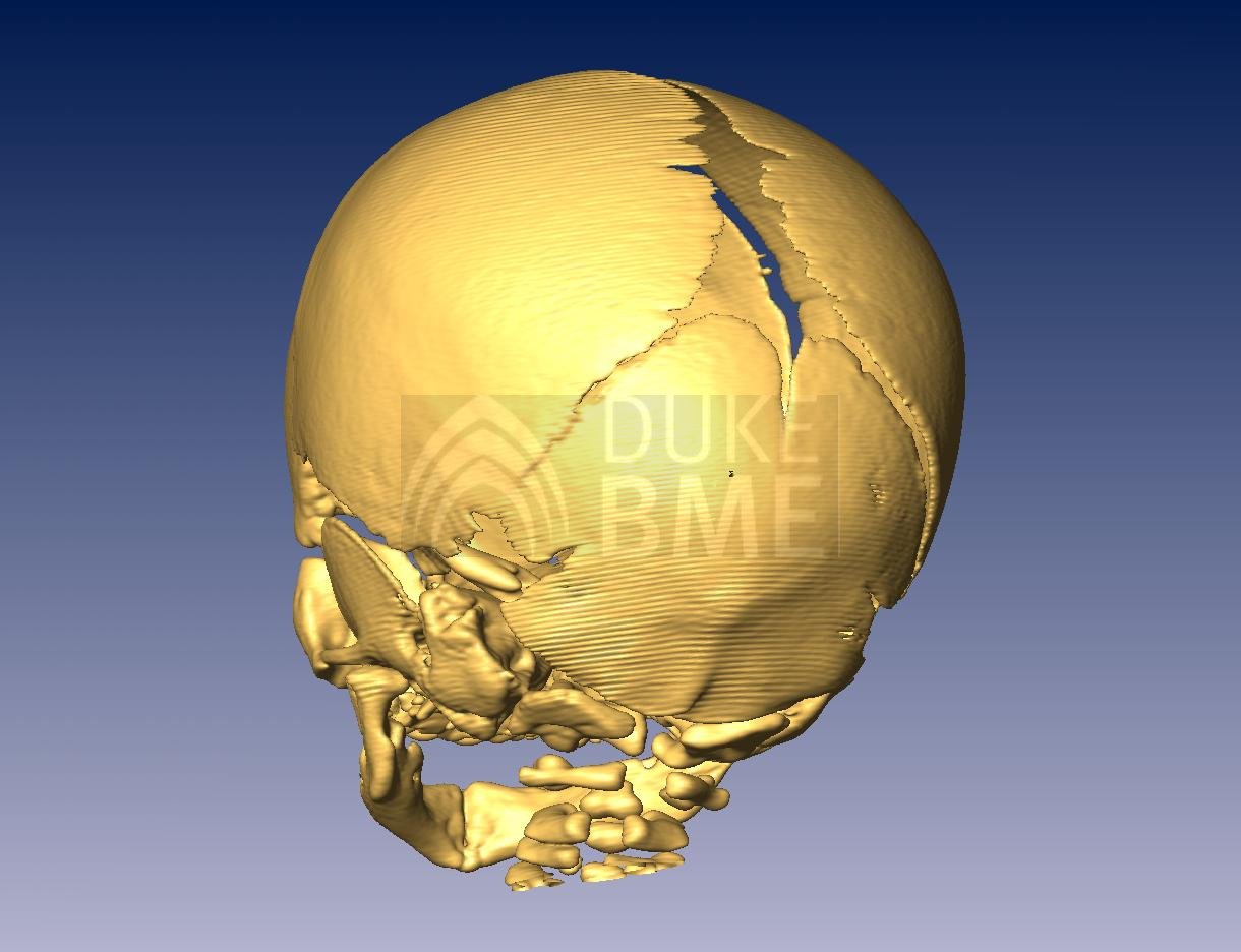
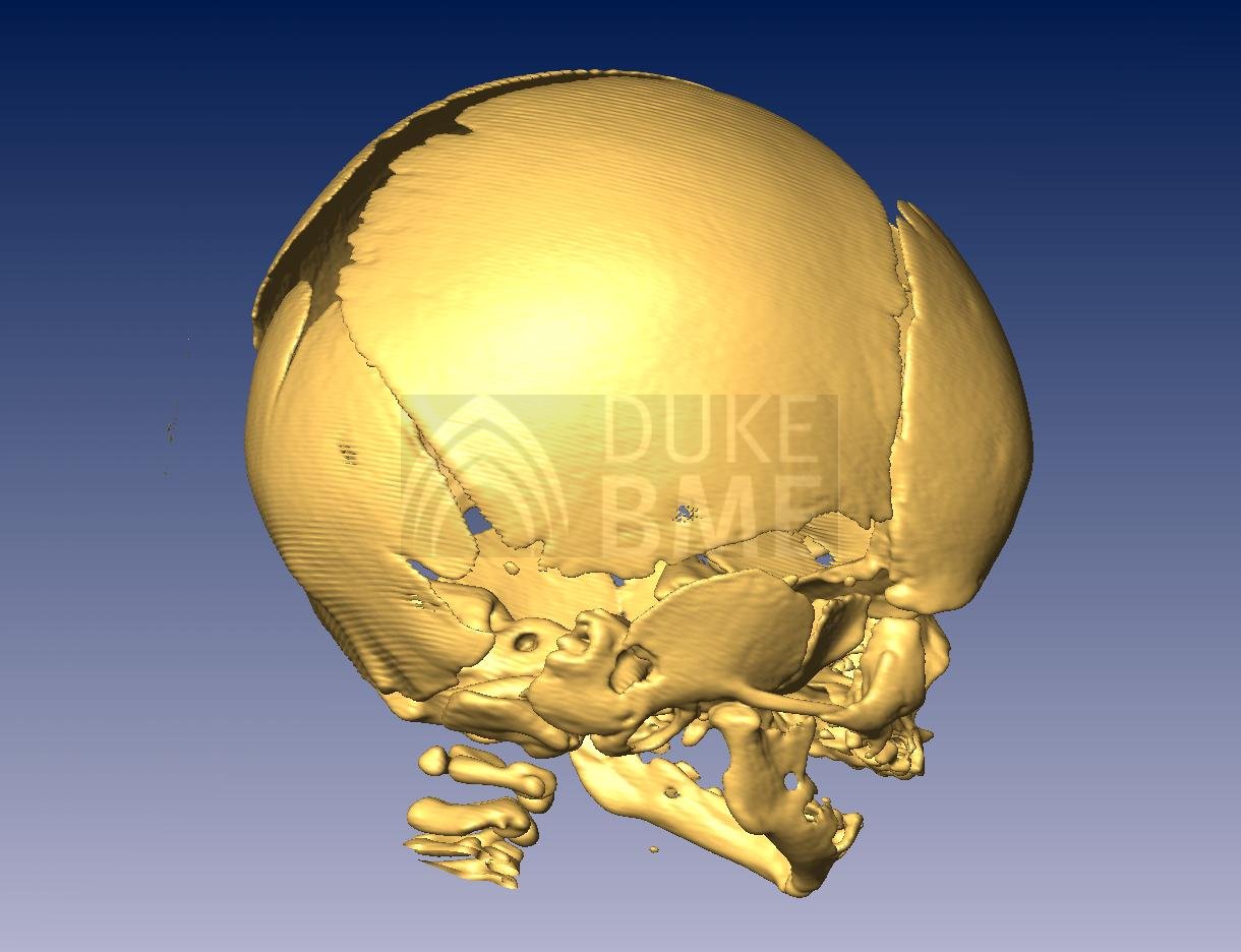
Below are examples of CT data rendered in AMIRA for visualization of the pediatric skull. The data are used for developing normative anatomical data for the skull and cervical spine. This information is important for identifying abnormal pediatric development and for the creation of computer models for injury research and for surgical management of deformities. Additionally, CT scans are being used to provide anthropometric information on how the head changes during the growth and development of the child.
Image
| Image
| Image
|